CTSA Logomark in front of red decorative line

Lung Cancer

Lung cancer is a common form of cancer that starts in the tissues of the lungs. There are multiple forms of the cancer, with each requiring unique treatment plans and prognoses. Approximately 75% of patients will have symptoms from locally advanced disease at their initial physician visit. The smoking rate in the US is 24% (45 million), with 90% of lung cancers thought to be smoking related. Smoking cessation is beneficial at any age, but much greater benefit is seen in those quitting at younger ages. The risk of lung cancer does not decline for many years following cessation, making screening very important.
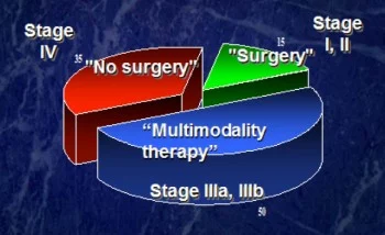
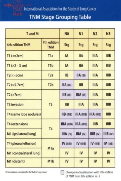
There are multiple kinds of lung cancers that we treat, including non-small cell lung cancers (NSCLC-87% of lung cancers ), which include adenocarcinoma, squamous cell carcinoma, and bronchioalveolar carcinoma, as well as neuroendocrine lung cancers, which include carcinoid, atypical carcinoid, large cell lung cancer, and small cell lung cancer (SCLC- 13% of lung cancers). The treatment a patient receives is determined by the pathology, or type of cancer they have been diagnosed with, as well as by the stage of the cancer. The lung cancer staging system is referred to as the TNM staging system. The “T” stands for tumor, and is determined by the size of the tumor, how many tumors there are, where the tumor is located, and if the tumor is growing into any nearby structures. The “N” stands for lymph nodes, and is determined by whether there is cancer found within them. It is also determined by which lymph nodes are involved. N1 lymph nodes are found within the same lung as the cancer. N2 lymph nodes are found in the mediastinum, which is the space in the chest between the two lungs. N3 lymph nodes are found in chest or lung on the side opposite the cancer. The “M” stands for metastatic disease, and is determined by whether the cancer has spread to any other parts of the body outside the lung such as the brain, liver, adrenal glands, bone, or the opposite lung. The final stage determines the treatment that is recommended. Stage I is usually treated with surgery alone. Stages II & III are usually treated with a combination of surgery, chemotherapy, and/or radiation therapy. Stage IV is usually treated with chemotherapy and /or radiation therapy alone.

Lung Cancer 5-Year Survivals

  • Stage IA = 75-80%
  • Stage IB = 60-75%
  • Stage IIA = 50%
  • Stage IIB = 40%
  • Stage IIIA = 25%
  • Stage IIIB = 10%
  • Stage IV = 5%
  • Overall SCLC (limited & extensive) = 6%
  • Overall NSCLC (all stages combined) = 15% (no change in 3 decades)

Usually a patient is seen by his or her physician for a physical examination. A chest X-ray or CT scan (computerized tomography) may be obtained if there are any concerning findings. Typically, a CT scan is more serviceable following such findings because it takes detailed pictures of the tissues inside the body. As you lie on the table, a CT scanner rotates around you while the table passes through the center of the scanner. During this time, pictures are taken that may show tumor, abnormal fluid, or swollen lymph nodes. Once an abnormality has been found, this requires further testing to determine the pathology. There are multiple tests that can be performed to obtain the diagnosis. Thoracentesis is where a long needle is used to take fluid from the chest cavity (pleural space) and the laboratory looks for cancer cells. Bronchoscopy is where a thin lighted tube (bronchoscope) is place through the nose or mouth, down the windpipe (trachea) to assess the air passages to the lungs. It allows sampling of cells within the airway. An ultrasound probe (endobronchial ultrasound – EBUS) along with a biopsy needle can also be used to take biopsies of the lymph nodes along the outside of the airway. Some patients require a CT guided needle biopsy, where the radiologist uses a long needle to directly biopsy the area of concern within the lung tissue. Sometimes it will even require a surgical procedure, such as VATS, mediastinoscopy, or thoracotomy (see Surgical Procedures), to finalize the diagnosis.

Once a patient has been diagnosed, staging is the next step. A PET scan (Positron Emission Tomography) will be obtained to see if there is any evidence of cancer in the lymph nodes or anywhere else in the body outside of the lung. You receive an injection of a small amount of radioactive sugar. A machine makes computerized pictures of the sugar being used by cells in the body. Cancer cells use sugar faster than normal cells, and the areas with cancer look brighter on the pictures. If there is concern for more advanced disease, an MRI of the brain might be ordered. This is a powerful magnet linked to a computer which makes detailed pictures of the tissues on a computer screen or film. A bone scan will sometimes be obtained to see if cancer has spread to the bones.

Once a patient has been diagnosed and staged, it is important to individualize the treatment based on the pathology, stage, patient’s performance status, other risk factors, and pulmonary function. Pulmonary function tests will be obtained to determine how much lung can be safely removed, without causing excessive shortness of breath to the patient after surgery. With full force, you exhale into a special instrument called a spirometer that measures how fast you exhale and how much air you can blow out. The spirometer is connected to a computer that records the measurements. The case will then be discussed at our multidisciplinary tumor board where an official recommendation can be made regarding treatment options. Once all the necessary testing is complete, and a tumor board recommendation has been made, you will discuss the results with your surgeon, and a plan will be tailored to your specific needs.